")
Herpes zoster ophthalmicus in HIV/AIDS
Related content
Herpes zoster is a common infection caused by the human herpes virus 3, the same virus that causes chickenpox. It is a member of herpes viridae, the same family as the herpes simplex virus, Epstein- Barr virus, and cytomegalovirus. Herpes zoster ophthalmicus occurs when a latent varicella zoster virus in the trigeminal ganglia involving the ophthalmic division of the nerve is reactivated. Of the three divisions of the fifth cranial nerve, the ophthalmic is involved 20 times more frequently than the other divisions.
Risk factors
Risk factors include the following:
- Decreasing immuno-competence
- Increasing age.
Immune suppression may be due to the human immunovirus (HIV) infection, malignancy, systemic lupus erythematosus, and the use of immunosuppressive agents. HIV positive patients have a 15-25 times greater prevalence of zoster compared to the general population.1 In the immuno-compromised patient, the dermatitis and ocular inflammatory disease are more prolonged and it is more difficult to prevent complications. Herpes zoster ophthalmicus may be the initial clinical manifestation of HIV infection.
The highest rise in prevalence, due to age, is in the fifth decade of life.
Extraocular manifestations of herpes zoster ophthalmicus
Infection and inflammation secondary to zoster can affect virtually all adnexal, ocular and orbital tissues.
Prodromal Stage
- Flu-like illness with fatigue, malaise, and low grade fever and chills that last up to one week before the rash over the forehead appears
- Pain: usually non-painful actions, like putting on a hat and combing hair may be very painful in about 60% of patients.
Rash
- Erythematous macules appear along the involved dermatome
- Over several days these progress into papules and vesicles, and later pustules, which rupture and crust, taking several weeks to heal
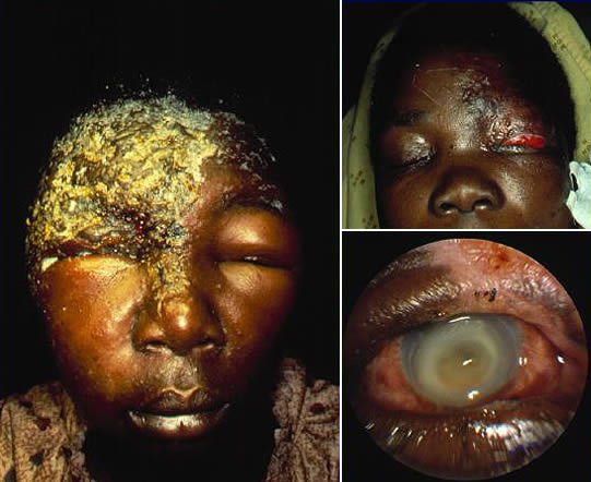
- HIV positive patients may have a generalized vesicular rash and become very ill one to two weeks after the onset of the disease, resulting in very severe visual impairment.
Ocular manifestations of herpes zoster ophthalmicus
The skin manifestations of herpes zoster ophthalmicus strictly ‘observes’ the midline with involvement of one or more branches of the ophthalmic division of the trigeminal nerve, namely the supraorbital, lacrimal, and nasociliary branches. Because the nasociliary branch innervates the globe, the most serious ocular involvement develops if this branch is affected. Classically, involvement of the tip of the nose (Hutchinson’s sign) has been thought to be a clinical predictor of ocular involvement.2 It is important to note that patients with a positive Hutchinson’s sign have twice the incidence of ocular involvement, but one third of patients without the sign develop ocular manifestations.
Eyelid
The eyelids are commonly involved in herpes zoster ophthalmicus.
- The majority of patients will have vesicular lesions on the eyelids that resolve with minimal scarring
- Patients may develop blepharitis. This can lead to secondary bacterial infection, eyelid scarring, marginal notching, loss of eyelashes, trichiasis and cicatricial entropion. Scarring and occlusion of the lacrimal puncta or canaliculi may occur
- Ptosis, secondary to oedema and inflammation may also occur.
Conjunctiva
Conjunctivitis is one of the most common complications of herpes zoster ophthalmicus. The conjunctiva is often injected and oedematous. This generally lasts for only one week. Secondary infection with Staphylococcus aureus may develop thereafter.
Sclera
Episcleritis or scleritis associated with herpes zoster may be either nodular or diffuse and can persist for months.
Cornea
Corneal complications occur in approximately 65% of cases with herpes zoster ophthalmicus.3 This can result in significant visual loss.
Symptoms are pain, photosensitivity and poor vision.
The clinical features of corneal disease in herpes zoster ophthalmicus may be due to:
- Direct viral infection
- Antigen – antibody reactions
- Delayed cell-mediated hypersensitivity reactions
- Neurotrophic damage.
Epithelial keratitis: The earliest manifestation of corneal involvement is punctate epithelial keratitis. Multiple, focal swollen lesions stain with fluorescein or rose bengal. These lesions contain live virus and may either resolve or progress into dendrites, presenting as early as one or two days after the initial rash, while dendrites often present after four to six days but can appear many weeks later. The dendrites appear as elevated plaques and consist of swollen epithelial cells. They form branching or ‘medusa-like’ patterns and have tapered ends in contrast to herpes simplex virus dendrites, which often have terminal bulbs. These dendrites also stain with fluorescein and rose bengal dyes. These epithelial lesions can lead to anterior stromal corneal infiltrates.
Stromal keratitis: This is an immune reaction to viral glycoprotein antigens deposited during the acute attack and possibly during late sub-clinical migration of the virus from the ganglion. Chronic stromal keratitis can lead to vascularization, corneal opacification, keratopathy, corneal thinning and astigmatism.
Uveal Tract
Anterior uveitis occurs frequently with herpes zoster ophthalmicus. The inflammation is generally mild and transient, frequently causing a mild elevation of intraocular pressure. Without timely and appropriate treatment the course of the disease may be prolonged and can lead to glaucoma and cataract.
Retina
The retinitis of herpes zoster ophthalmicus is often associated with anterior uveitis. It presents as necrotizing retinitis with haemorrhages and exudates, posterior vascular occlusions and optic neuritis.These lesions begin from the retinal periphery. The vision deteriorates rapidly as the disease progresses.
Post-Herpetic Neuralgia (PHN) and Post -Herpetic Itch (PHI)
Pain and itching, late in the disease, are both acute and more common in HZO than in any other form of zoster. PHN is described as constant boring pain, sudden transient sharp pain, or pain elicited by usually non-painful stimuli. The mechanisms of PHN and PHI are not well understood but appear to be related to loss of peripheral sensory neurons.
Complications
- Corneal neovascularization and scarring resulting in poor vision.
- Neurotrophic ulcer with perforation.
- Secondary bacterial or fungal infection.
- Secondary glaucoma from uveitis or steroid treatment.
- Necrotizing interstitial keratitis.
- Post-herpetic neuralgia.
- Vision loss from optic neuritis or chorioretinitis.
Management of herpes zoster ophthalmicus
Herpes zoster ophthalmicus can be successfully managed by simultaneously combining systemic antivirals and tricyclic antidepressants to inhibit the infectious – inflammatory component and the pain. Antiviral agents may decrease the severity and duration of symptoms, if given early in the course of the illness.
Oral acyclovir therapy for herpes zoster ophthalmicus is found to:
- Reduce viral shedding from vesicular skin lesions
- Decrease systemic dissemination of virus
- Reduce the incidence and severity of the most common ocular complications such as dendritic keratitis, stromal keratitis and uveitis.4
Antibiotics should be used if secondary bacterial infection of the vesicles has occurred. Unfortunately, oral acyclovir has little effect on the incidence, severity, or duration of post-herpetic neuralgia.
Steroid eye drops may be helpful for HZO, but they can be disastrous for herpes simplex keratitis (Table 1). If secondary impetigo is present, a suitable anti-staphylococcal antibiotic should be given, and the patient should be considered for hospital admission because of facial cellulitis. If a patient complains of severe pain at any point at or beyond the appearance of crusted vesicles, assume that post-herpetic neuralgia has developed. It requires aggressive management. Narcotic analgesics may be used. Capsaicin cream may be applied topically to the affected areas six hourly.
Tricyclic antidepressants (e.g., amitriptyline) are often helpful in post-herpetic neuralgia; they should be initiated at low doses and increased as appropriate.
Table 1. Suggested management protocol
| Infection | Treatment |
|---|---|
| ‘Shingles’ | Acyclovir (Zovirax) 800mg orally five times daily for 7 to 10 days |
| Skin | Apply cool compresses |
| Blepharitis/ Conjunctivitis | Topical lubrication. Topical antibiotic. |
| Epithelial keratitis | Debridement or none |
| Stromal keratitis | Topical steroids |
| Neurotrophic keratitis | Topical lubrication Topical antibiotics Tissue adhesives and protective contact lenses to prevent perforation |
| Uveitis | Topical steroids Oral steroids Oral acyclovir |
| Scleritis/ Episcleritis | Topical non-steroidal anti-inflammatory agents and/or steroids |
| Acute retinal necrosis/Progressive outer retinal necrosis | IV acyclovir (1,500 mg per m2 per day divided into three doses) for 7 to 10 days, followed by oral necrosis – acyclovir ( 800 mg orally five times daily) for 14 weeks |
Conclusion
Patients and the general population should be instructed regarding the importance of early presentation and careful compliance in treatment as well as the importance of regular follow-up. One obstacle that confronts eye care workers in the developing world is the cost of the drugs which affects compliance, as the patients are poor.
References
1 Pavan-Langston D. Clinical manifestations and therapy of herpes zoster ophthalmicus. Comp Ophthalm Update 2002; 3: 217.
2 Sheikh S, Ta C N. Evaluation and management of herpes zoster ophthalmicus. Am Fam Physician 2002; 66: 1723-1730.
3 Pavan-Langston D. Clinical manifestations and therapy of herpes zoster ophthalmicus. Comp Ophthalm Update 2002; 3: 219.
4 American Academy of Ophthalmology. External Disease and Cornea. Basic Clinical Science Course Manual 1998-1999; 8: 154.