")
Gram stain
Related content
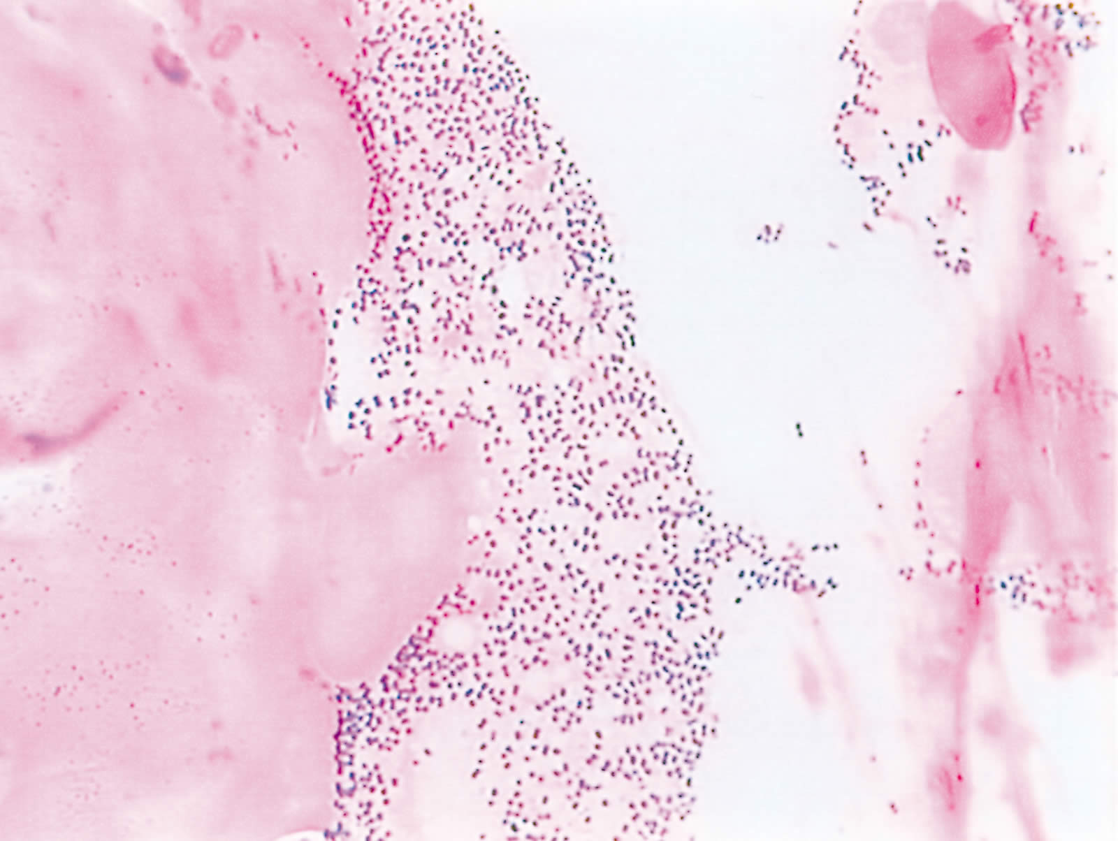
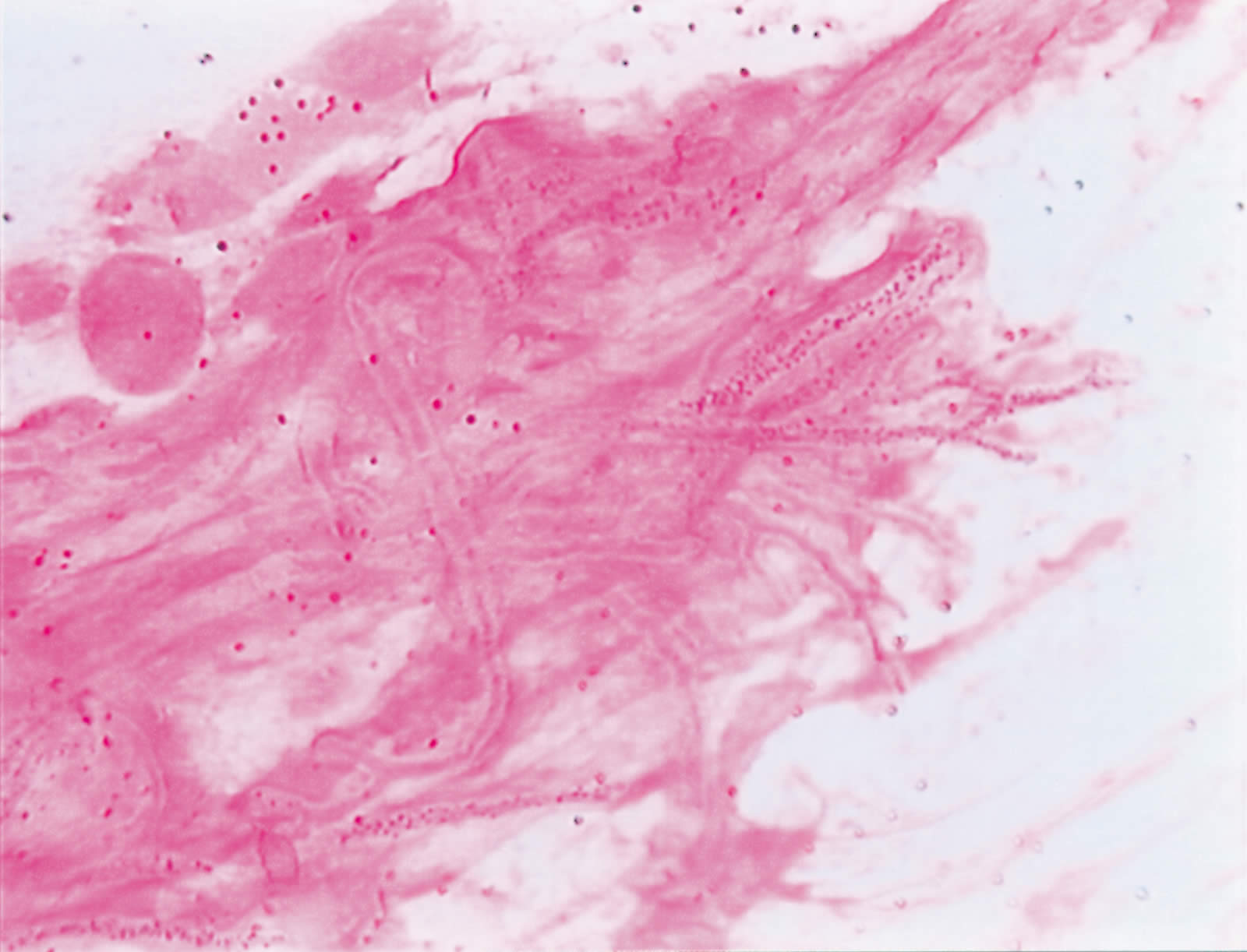
This is by far the most important staining method in bacteriology. It is a staining technique which is employed for the diagnostic identification of a wide variety of organisms. The mechanism of the Gram stain is not fully understood beyond the identifiable differences in cell wall characteristics between those organisms classified as ‘Gram +ve’ and those classified as ‘Gram -ve’. The Gram +ve organisms are able to retain basic dyes at a higher concentration than the Gram -ve species. Probably, the most important difference is in the permeability of the cell wall during the staining process.
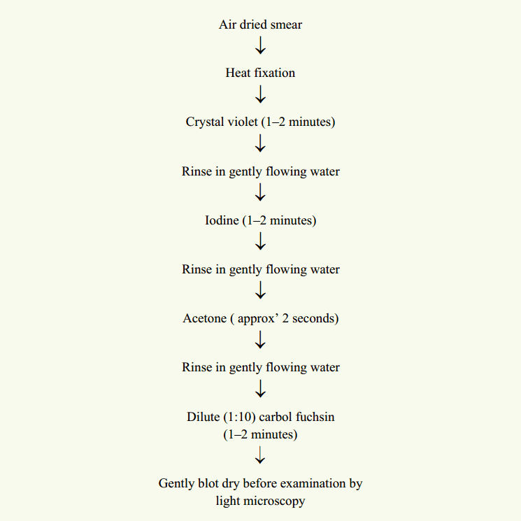
Following staining with crystal violet and treatment with iodine, a dye-iodine complex is formed within the cell. This is insoluble in water but moderately soluble in acetone (or alcohol) which is used as a decolouriser. Under the influence of the decolouriser the dye-iodine complex (blue/black in colour) is retained by the Gram +ve group of organisms but flows freely from the Gram -ve group. Presumably, this is due to the former having a less permeable cell wall. The Gram -ve group can now assume the colour of the chosen counter-stain to distinguish between the two groups.