")
A low-cost, slit lamp-based video-photodocumentation system
Related content
Editor’s note
We are aware there are many different methods of capturing images for teaching and other purposes, most frequently using the existing optics of the slit lamp. Dr Nizvi has sent us details of his own method. We would emphasise the need for background illumination in the majority of images.
Introduction
Video display of the slit lamp view of the eye is useful for various purposes in ophthalmology, such as teaching, record keeping, and teleophthalmology. However, commercially available photo slit lamps are quite expensive. In this short article, I describe a low-cost system providing fairly good quality video images using an ordinary camcorder. I have been using this system for teaching community eye health workers and educating patients for ten years.
Basic principle
This can be considered in two parts.
- Changes in the optics of the camcorder such that its focus is adjusted at a distance equal to the working distance of the biomicroscope. The magnification changes accordingly.
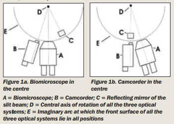
- Attachment of the camcorder besides the biomicroscope such that the optical axes of the camcorder, the biomicroscope, and the slit beam are in the same plane, meet at the same point, and share the same rotational axis in all positions. During normal working position, the camcorder shows the view at some angle with that of the examiner’s view (Figure 1a). If an exactly frontal view is desired, or gonioscopy or slit lamp ophthalmoscopy needs to be done, the camcorder has to be shifted to the centre (Figure 1b), and the examiner looks through the viewfinder or the monitor.
Any camcorder can be used provided that manual focus and exposure options are available. The smaller the camcorder in size the less obtrusive it will be. In this study a JVC GR-DV1 (Victor Company of Japan, Limited) was used. The slit lamp was manufactured by Haag-Streit.
Part 1 of the principle is achieved by fixing, in front of the camcorder’s optics, a +9.0 dioptre lens taken from the trial frame, as the optics of a camcorder resembles that of the surgical microscope with the objective lens removed.
To achieve part 2, a metal adaptor was made by the author.
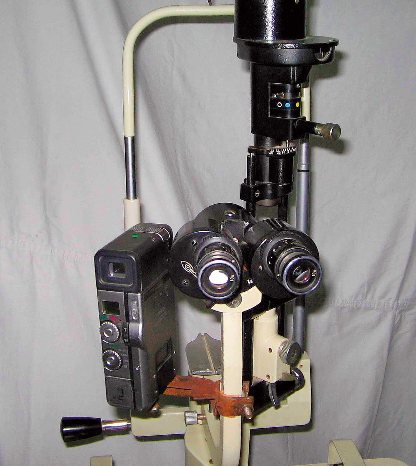
The camcorder is fixed to it by means of a screw into the tripod mounting socket. The adaptor is fixed to the arm of the biomicroscope carriage (Figure 2). The screws are not fully tightened yet.
The camcorder is shifted in the centre and the focusing rod provided with the slit lamp is inserted in place. The manual focus mode is set and the slit beam is turned on. Fine adjustments are made to bring the slit beam image in the centre of the viewfinder, and the screws are tightened in this position. Next, the fine focusing is done and the rod is removed. A source for diffuse light (Canon video camcorder flash) is attached beside the reflection mirror of the slit beam for background illumination. (Any diffuse light source can be used). The video-out lead is attached to an ordinary colour television. (The video-out lead can also be attached to a video capture card in a personal computer).
Discussion
Images obtained by this system are of reasonably good quality. The use of a photographic quality objective lens can greatly improve image quality, and with some basic knowledge of photography and some experience, most of the clinical conditions can be documented clearly. The system has the extra advantage of wide range zoom magnification with an ordinary slit lamp. It also obviates the need of a separate video recorder (VCR) for recording. The camcorder can be used for general purpose at any time just by loosening a screw and removing the objective. A professional manufacturer can design an adjustable, universal adapter that can fix different camcorders to slit lamps of various models.